New market-ready X-ray camera developed for colour imaging
GOTT funded STFC to develop a market-ready X-ray camera, enabling colour imaging and access to the chemical composition and microstructure of samples.

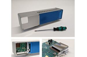
The picture on the left shows the HEXITEC-MHz camera system.
Background
X-rays are widely used in various fields such as medicine, material science and astronomy.
However, conventional X-ray detectors do not record the energy of incoming X-ray photons, and thus produce black-and-white images giving little information about a sample.
The Science and Technology Facilities Council (STFC) has been developing a market-ready colour X-ray camera system for over a decade. This innovative technology enhances imaging capabilities by measuring the energy of X-ray photons that are transmitted, absorbed and scattered by a sample.
Many colour images are produced each representing a narrow energy window (1keV), which are then compiled together to make detailed, high-resolution X-ray images with additional colour information, providing access to the sample’s chemical composition and microstructure.
The Knowledge Asset solution
Initially, STFC’s Technology Department developed High Energy X-ray Imaging Technology (HEXITEC). While HEXITEC was successfully used in various areas, its applications in many industry-relevant techniques were limited.
Funding awarded by the Government Office for Technology Transfer (GOTT) was used to support the development of the next generation of this technology, the HEXITEC-MHz camera.
The new HEXITEC-MHz runs 100 times faster than the original system allowing high-fidelity colour X-ray imaging to be applied at greater fluxes. This has opened new potential application areas such as colour CT imaging for material science and non-destructive testing. The new camera system will be more user-friendly and ready for the market, after hardware development and data processing optimisation.
The HEXITEC-MHz camera will provide users with information about the composition of a sample, including its structure, what materials are present, and their quantities.
Who will this help?
- Medical imaging: It will identify and classify different types of tissues during biopsies, providing clearer and more detailed images for better diagnosis and patient treatment.
- Security: It will enhance the ability to detect and identify objects in security screenings by probing the molecular structure and components of a sample to reduce false alarm rates.
- Industrial inspection: It will reduce the scan times from hours to minutes with added colour imaging, improving the accuracy of inspections in manufacturing and other industries.
Funding awards
STFC was awarded £202,812 in the ‘Extend’ band of the Knowledge Asset Grant Fund in August 2022.
GOTT’s role
GOTT provided grant funding to the project.
Matt Wilson, STFC Technology Department’s Detector Development Group Leader, said:
The Knowledge Asset Grant Fund has been a valuable and timely way to increase the technology readiness level of our unique HEXITEC-MHz X-ray detector system. GOTT’s team helped make the application and grant management process simple and has given us a focus to target the best routes to market for our technology.
The development team from across the Technology Department has been able to demonstrate continuous MHz data collection and processing on the fly, which is key to making high speed colour X-ray imaging seamless for the user. We are now in a position to showcase our detector with our collaborators in science and industry.
Early results
The new X-ray camera system has been in testing and shown promising early results. HEXITEC-MHz was installed at Diamond Light Source, the UK’s national synchrotron science facility, to demonstrate the feasibility of a novel X-ray optical technique, called ptychography, in imaging small objects that are difficult to see through traditional microscopes.
The team are now preparing to take the product to industry.