Guidance for diagnostic investigation of acute mortality with skin scald in pigs – for vets
Published 27 January 2025

© Crown copyright 2025
This publication is licensed under the terms of the Open Government Licence v3.0 except where otherwise stated. To view this licence, visit nationalarchives.gov.uk/doc/open-government-licence/version/3 or write to the Information Policy Team, The National Archives, Kew, London TW9 4DU, or email: psi@nationalarchives.gov.uk.
Where we have identified any third party copyright information you will need to obtain permission from the copyright holders concerned.
This publication is available at https://www.gov.uk/government/publications/guidance-for-diagnostic-investigation-of-acute-mortality-with-skin-scald-in-pigs-for-vets/guidance-for-diagnostic-investigation-of-acute-mortality-with-skin-scald-in-pigs-for-vets
1. Background information
Since 2010, APHA has investigated, with private veterinary surgeons, eight incidents of acute, transient mortality with skin scald in growing pigs. Differentials have included septicaemia, water deprivation, magnesium toxicity, acute zinc toxicity and adverse tiamulin reaction, however no diagnoses were confirmed in any of these cases.
Investigations have involved collection of detailed epidemiological and clinical history, farm visits with examination of live pigs and post-mortem examinations with bacteriology and histopathology. Some cases have included biochemistry and feed analysis for mycotoxins and nutritional imbalances.
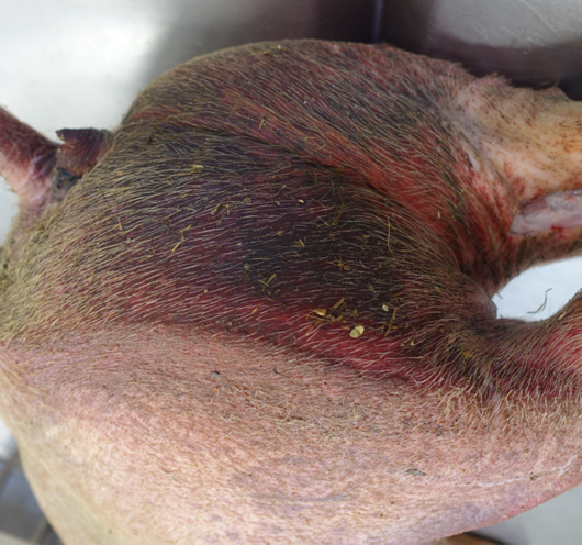
All cases occurred between August and October on solid-floored indoor straw units. Deaths involved three to more than 100 pigs over 24 hours. In most incidents, live pigs have appeared ‘off colour’, agitated and/or had reddened skin. The surviving pigs in each of these cases recovered fully. Deaths and clinical signs were restricted to pigs in a small number of pens in five cases. The gross pathology in all cases involved severe, mainly ventral, skin lesions resembling scald (Figure 1), with visceral congestion and haemorrhages in seven cases, without lesions typically associated with swine fevers
Histopathology in all cases revealed changes consistent with a severe, acute, irritant, contact dermatitis. No bacterial pathogens were isolated from a range of systemic sites. Biochemistry was unremarkable, in particular, urine or blood magnesium was not raised. No feed imbalances have been detected. Whole Genome Sequencing of E. coli from one case did not detect genes for urease production. In another case, deoxynivalenol was detected in feed but not at a clinically significant concentration.
Three incidents were preceded by a recent interruption in feed supply and/or new feed delivery. Hypotheses for this clinical presentation include mycotoxicosis due to consumption of mouldy feed dislodged during feed bin refilling or accumulation of a feed fraction residue. A further four incidents had a very recent history of whole-group, in-water oral antibiotic use, but tiamulin treatment (a potential initiating factor for this clinical presentation) was not described in any case.
APHA wishes to hear of similar cases and can provide assistance with diagnostic investigations, by advancing understanding of possible risk factors and ruling out differential diagnoses.
2. Suggested investigation in similar cases
Before proceeding with diagnostic investigations, the attending vet should ensure there is no reason to suspect notifiable disease, in particular, African or Classical swine fever. This may be achieved through consideration of the clinical and epidemiological history, as well as examination of live pigs and post-mortem examinations. Images of the clinical signs and pathology related to African swine fever are here. Suspect notifiable animal disease must be reported to Defra Rural Services Helpline on 03000 200 301 in England. In Wales contact 0300 303 8268 and in Scotland contact your local Field Services Office.
2.1 Collection of epidemiological and clinical history
In addition to standard clinical and epidemiological details:
- Record timeline and pattern of disease progression across pens;
- Determine mortality and morbidity;
- Establish whether water and/or feed supply may have been interrupted;
- Explore interruptions or changes in feed, including when the feed bin(s) was last refilled;
- Understand any recent antibiotic use, in particular, tiamulin use;
- Discuss changes in management, bedding, etc.
2.2 Clinical examination of live pigs
Perform thorough clinical examinations of affected pigs. Record rectal temperatures and take photos and videos. Collect clotted, EDTA and heparin blood for biochemistry from affected pigs which appear ‘off colour’, agitated and/or have reddened skin.
2.3 Postmortem examinations
Vets can submit carcasses to an APHA veterinary investigation centre or a partner postmortem provider for postmortem examination and diagnostic testing. In Scotland, contact SRUC. Where on-farm postmortem examinations are undertaken:
- Record post-mortem findings and take photos where appropriate;
- Collect and fix (as quickly as possible) tissues for histopathology from all major organs (liver, lung, spleen, heart, lymph node, kidney, pancreas, skeletal muscles) and from any lesions. Collect and fix several sections of affected and unaffected skin.
- Using aseptic methods (as far as possible), take charcoal swabs from systemic sites for bacteriology (e.g. meninges, liver, lungs and spleen);
- Fix brain (where possible) for histopathology;
- Collect fresh kidney and liver samples to store frozen;
- Collect urine (where present), bile (to freeze) and stomach contents. Use a needle to collect aqueous humour from the anterior chamber of the eyes.
2.4 Collection of feed samples
Collect feed samples (>2kg) as early as possible in the course of disease from feed hoppers in the most affected pens for mycotoxin analysis, including citronin and, if magnesium is raised in the pigs, for magnesium analysis.
2.5 For further assistance with disease investigations
- Contact your nearest APHA Veterinary Investigation Centre; or
- Email the Veterinary Lead of the Pig Expert Group.